Figure 1: Osteosarcoma in adult female rat (Zinnia).
Case history and photos
History
A 4 to 6-month old spayed female rescue rat weighing 355 grams.
She was being treated for respiratory illness on maintenance dosing of Baytril and doxycycline.
Clinical Signs
About 12 months after adoption (16 to 18 months old) a bony tumor approximately 0.5 cm in diameter was found on her left tibia.
Clinical signs also included limping and gradual weight loss. Within 4 to 5 months the mass was the size of a golf ball.
Testing and Initial Diagnostics
A fine needle aspiration of the nodules revealed mononuclear cells typical of neoplasia. Initial diagnosis was of an unknown slow-growing inoperable tumor. Post postmortem histopathology determined the mass was an osteosarcoma.
Treatment
Due to several factors, including Zinnia’s age, whether there were possible metastases and her ongoing treatment for respiratory disease, it was decided not to pursue amputation of the left hind leg with tumor. Palliative care was thought to be the best course of action.
Pain management consisted initially of ibuprofen (15mg/lb), then Metacam (2 drops daily), then prednisone, and finally Tylenol (90mg/lb). Prednisone was continued to minimize growth and provide comfort. The Baytril and doxycycline were continued.
In an effort to reverse/stabilize her weight loss her dietary intake was increased with moderate amounts of Ensure, avocado, scrambled eggs, and cooked oatmeal.
For safety and ease she was placed, along with two female cage mates, in a one-story cage with smooth ramp to a low shelf.
Outcome
Due to progression of the tumor and advancing pneumonia Zinnia was euthanized. The time between the owner noticing the leg mass and euthanasia was approximately 5 months. Her weight at death was 300 grams.
Histopathology
Summary:
Pathology comment: features of the mass lesion of the left hind limb consistent with a diagnosis of malignant osteosarcoma.
Report:
The mass from the left hind limb consists of pleomorphic neoplastic plump spindle-shaped to polygonal osteoblasts forming sheets and broad interlacing bundles, surrounding coalescing lakes of osteoid which is variable well mineralized, and with foci of osteocartilaginous metaplasia. Tumor cells have moderate anisocytosis and anisokaryosis, mitotic figures average one per high power field.
Photos
 The left photo is of Zinnia showing a normal left hind limb one year before the mass began to develop. The middle photo was taken one year later directly after a fine needle aspiration was performed. The last photo shows mass size during that same month. |
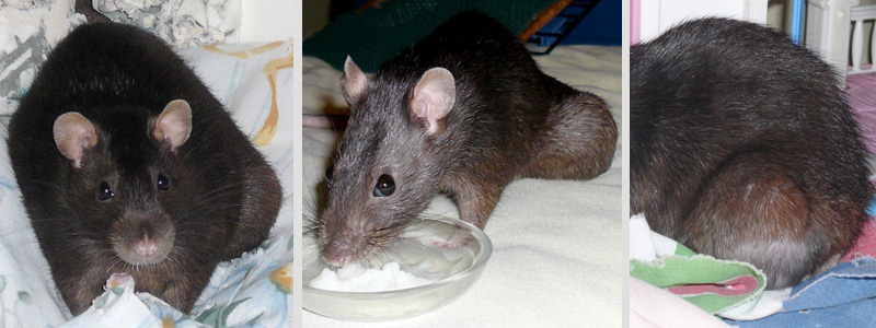 These three photos show continued growth of the mass one month following fine needle aspiration. |
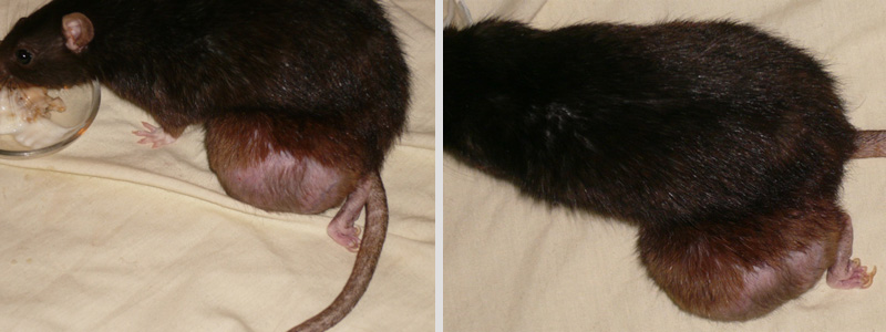 These two photos were taken prior to euthanasia, and approximately 5 months from the initial diagnosis. The mass on the left hind limb at this time was approximately the size of a golf ball. Histopathology confirmed osteosarcoma. |
Veterinarian- Paul Palmetier, DVM
Pathologist- Shane Stiver, DVM
Case history and Photos courtesy of Carolyn Sekela RNC-e.

