Figure 3: Pituitary tumour overview
The following photographs provide a dorsal view into the (opened) skull cavity of adult male and female rats who were found to have a pituitary tumour during necropsy. All of these rats were symptomatic for varying periods of time before euthanasia was required due to the pituitary symptoms (and in two cases comorbidity).
Note that the ages mentioned below are sometimes estimates, due to the rats in question being “rescue rats” with uncertain birthdates. Also note that it can be difficult to determine the exact age at which pituitary symptoms first appear, since the symptoms can vary, not every rat displays them in the same order or to the same degree, and especially the early symptoms may be overlooked altogether, or confused for symptoms of comorbid conditions.
Unless otherwise mentioned, all rats received veterinary treatment (medication) for an at that point in time presumed pituitary tumour. Unfortunately, there were only two cases in which it seems to have had some positive effect.
The third photograph of row 3 shows approximately how the rats are oriented (with two exceptions) and how the opening in the skull was created. The cranium (labelled “S”) was cut with scissors, starting at the back/base of the skull via the foramen magnum and continuing as far left and right as possible along its outer edges; then flipped towards the nose, displaying the inner/ventral surface of the bone. After severing the optic nerves (indicated by the blue arrowheads) between the eyes and the brain (labelled “B”), the latter was gently pried out of the skull cavity and flipped towards the spine, also displaying its ventral surface. While doing this the pituitary tumour (indicated by the white arrow) remains in place, as it is attached to the “sella turcica”, an indentation of the sphenoid bone beneath it. Although the rats in these photographs are of varying bodyweight, the opening created in their skulls is of roughly the same size, allowing for a rough comparison of the size of the pituitary tumours found (several of them were also measured, either in situ or after excision).
All pituitary tumours were found to be “firm masses”, unless stated otherwise.
Necropsy Pictorial
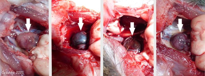 Row 01:These photographs are of pituitary tumours (indicated by white arrows) found in four adult male rats. In the first two cases the symptoms presented around 1.5 years of age and progressed quite rapidly, requiring euthanasia within 1-2 months. The 1st pituitary tumour was appr. 1 x 0.7cm in situ, with a possible relatively small caudal “blood blister”. The 2nd pituitary tumour was appr. 1cm in diameter; there was a relatively small ventral “firm” portion and a large dorsal “blood blister” that “deflated” when squeezed gently. The male in the third photograph required euthanasia within 2-3 months, after symptoms presented at 2 years and 8 months of age. (No medical treatment.) His pituitary tumour was appr. 0.5cm in diameter. The male in the fourth photograph is thought to have shown symptoms for possibly 10 months, starting at 1 year and 9 months of age. His pituitary tumour was appr. 0.8 x 0.5cm in situ and consisted of a larger (cranial) and smaller (caudal) part, with an additional tiny spherical part on top of the latter. |
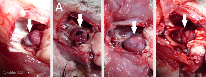 Row 02: These photographs are of pituitary tumours (indicated by white arrows) found in four adult female rats. In all of these cases the symptoms progressed quite rapidly, requiring euthanasia within 1-2 months (in the first case within 2 weeks). The first three rats presented with symptoms at or just under 1.5 years of age; the fourth rat at a little over 2 years of age. Note that in the first and final photograph the pituitary tumour consists of two parts, with the white arrow indicating the delineation between the two parts. The 2nd pituitary tumour was under 1cm in diameter and showed bleeding on its dorsal surface. The 4th pituitary tumour was appr. 1 x 0.5cm in situ. Interestingly, the 3rd pituitary tumour was found to contain a central cavity when cut longitudinally in situ. |
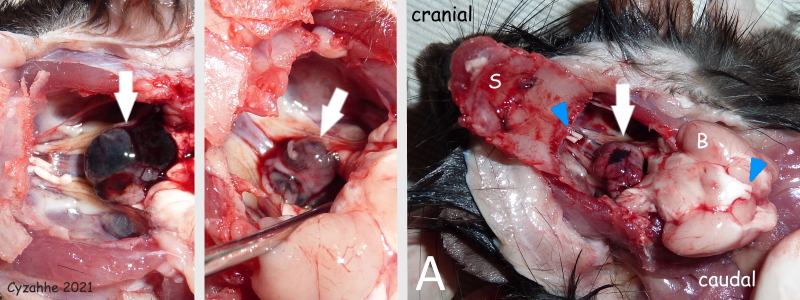 Row 03: The first two photographs in this row are of pituitary tumours (indicated by white arrows) found in two adult female rats who required euthanasia within 2-3 months, after symptoms presented at respectively slightly under 2 years, and 1 year and 9 months of age. The 1st pituitary tumour was accidentally nicked during the necropsy causing bleeding; the apparent “blood blister” was not examined further. The 2nd pituitary tumour was a little over 0.5cm in diameter. The third photograph is of the female rat labelled “A” in row 2. It shows approximately how the rats are oriented (with two exceptions) and how the opening in the skull was created. The cranium (labelled “S”) and brain (labelled “B”) have been flipped, revealing their ventral surfaces. The severed optic nerves (this is required to flip the brain) are indicated by blue arrowheads. |
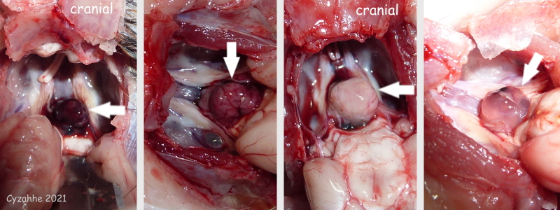 Row 04: These photographs are of pituitary tumours (indicated by white arrows) found in four adult female rats. (Note that the orientation of the rats in the first and third photograph is different, with the nose pointing upwards.) In all of these cases the symptoms progressed relatively slowly. In the first two cases symptoms were present for around 6 months before euthanasia was required, starting at respectively 2 years and 4 months, and 1 year and 3 months of age. The 1st pituitary tumour was appr. 0.3cm in diameter (no medical treatment); the 2nd appr. 0.7cm. The final two cases are thought to have shown symptoms for respectively well over a year to almost 1.5 years. In both cases the symptoms started at or just over 1.5 years of age. The 3rd pituitary tumour was appr. 0.7cm in diameter and strikingly pale. This female showed a possible slight brief improvement on oral prednisolone. The 4th pituitary tumour was appr. 0.8cm in diameter and appeared almost see-through; it was unclear whether it consisted of fluid or “mushy tissue”. Over the course of 1.5 years this female showed three “episodes” of sudden loss of strength, coordination, disorientation and lethargy. During the first episode she was given oral prednisolone (continued for 20 days) and made a full recovery by the following morning. During the second episode she received a single dexamethasone injection, followed up by oral prednisolone (for 1 month) and oral cabergoline (until euthanasia); she showed improvement in strength and manual dexterity the following day. During the third episode she was given oral prednisolone (until euthanasia), but did not show clear improvement. Overall, she showed slow but consistent decline. |
Graph Pictorial
The graph below depicts each rat’s age of onset of clinical signs/symptoms, duration, age at time of death.

|
Necropsies, photos, and descriptions by Cyzahhe
Edited by Karen Grant RN

