Urolithiasis and abdominal abscesses in male rat (Gambit).
Case History
An 11-month-old castrated semi-wild male rat. Clinical signs of illness at the time of euthanasia: urolithiasis, abdominal “lumps”, found in the middle of the night in severe pain and lethargic. Weight: 612 grams.
Necropsy Pictorial
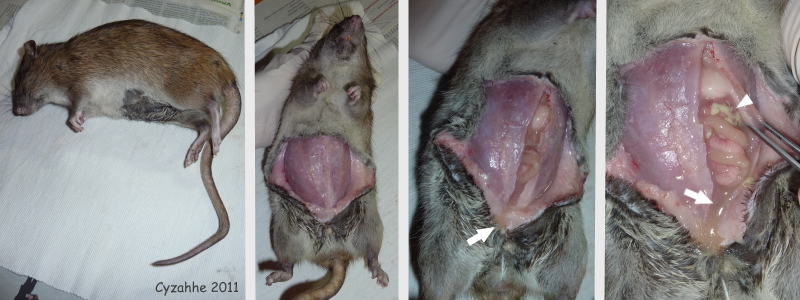 Row 01: The first photograph shows Gambit 2.5 hours post-mortem (unrefrigerated). As can be seen by the bends in his tail, rigor mortis has already set in. In the subsequent photographs the abdominal skin has been incised and peeled to the sides, after which the abdominal wall was incised. (Note that in semi-wild rats the fur and abdominal musculature is noticeably thicker than in fully domesticated rats.) Yellowish watery foul-smelling fluid immediately oozed from the abdomen (indicated by white arrows), indicating ascites, and greenish-yellow pus was visible (indicated by a white arrowhead). |
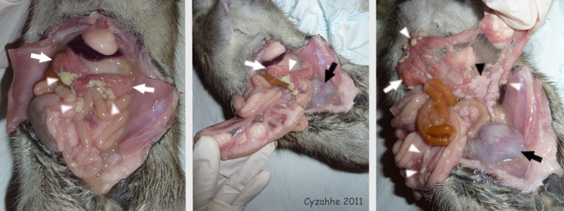 Row 02: In these photographs the abdominal wall has been peeled to the sides as well, allowing a full view of the in situ intestinal tract (first photograph). Once again note the greenish-yellow pus (indicated by white arrowheads), as well as the irritated-looking red spotting of parts of the mesentery (indicated by white arrows). In the second and third photograph parts of the intestinal tract have been pulled out of the abdominal cavity. The pancreas can be seen (indicated by a black arrowhead) and appears normal. The abdominal “lumps” are indicated by black arrows. |
 Row 03: In the first photograph the connection between the esophagus and stomach (indicated by a white arrowhead) has been severed, allowing for its removal from the abdominal cavity, along with the majority of the intestinal tract. Note that the spleen (indicated by a black arrowhead) is of normal size and shape. The second photograph is a close-up of the “lumps” (indicated by black arrows) adhered to the intestinal tract. The third photograph shows them from another angle. There were three semi-joined hard masses, surrounded by whitish fascia, with greenish-yellow areas showing through from inside. At this point it was still unclear whether the “lumps” were tumours and/or abscesses, and from which organ they stemmed. |
 Row 04: The first two photographs show different angles of the abdominal “lumps” (indicated by black arrows). Note the greenish-yellow areas showing through from inside (indicated by black arrowheads). The second photograph shows the irritated-looking red-spotted small bladder (indicated by a white arrow). In the third photograph the intestinal tract has been removed from the abdominal cavity and the “lumps” have been incised and (partially) expressed, resulting in the oozing out of foul-smelling greenish-yellow pus. When the largest “lump” was rinsed out with water only a whitish fascia sac remained, at which point the conclusion was drawn that they must be abscesses. It remains unclear from which organ they stemmed. |
 Row 05: The first photograph shows the liver after removal from the abdominal cavity. It has been incised longitudinally (indicated by a black arrow), as well as transversely (indicated by white arrows). The second photograph shows one of the kidneys after removal from the abdominal cavity. Note the tiny adrenal gland (indicated by a black arrowhead), as well as that both are embedded in adipose tissue (indicated by white arrowheads). In the third photograph the kidney has been incised longitudinally. Both organs appear normal. |
Histopathology
A necropsy is performed by Gambit’s caretaker 2.5 hours post-mortem (unrefrigerated). Tissue samples are collected and placed in formalin. Histopathology is performed a few weeks later. (Unfortunately, bacterial analysis could not be performed on the pus inside the lumps due to the formalin.)
MACROSCOPIC
Parts of the intestinal tract have adhered. Multifocal purulent sites surrounded by connective tissue. One site is at least 2cm in diameter.
MICROSCOPIC
Necropurulent sites with multifocal accumulations of bacteria, surrounded by thick connective tissue. Purulent sites within the connective tissue. An adjacent part of the intestinal tract has some plasma cells and heterophile granulocytes in its lamina propria. Lymphoplasma cellular infiltrates are present in other parts, in the direction of the serosa. Locally there is also some angiofibroblastic tissue.
CONCLUSION
Multifocal abscesses in and around the serosa of the intestine. No indication of neoplasia.
Note:
Gambit’s entire case history can be found in the Health section of Rat Guide in the article on Urolithiasis as Fig:3 Urolithiasis in 8-month-old neutered male rat. Multifocal abdominal abscesses identified in post mortem histopathology
Necropsy and photos by Cyzahhe
Histopathology by Utrecht University, Veterinary Pathology Diagnostic Centre (VPDC)
Edited by Karen Grant RN

