Figure 6: Vaginal sarcoma in a 26-month-old female rat (Jango).
Case history and photos
History
Jango was a 26-month-old Russian Blue Dumbo eared intact female rat, weighing 356 grams. She gave birth to one healthy litter, with no complications, and did not have any previous health issues.
Clinical Signs
The owner observed abnormal protrusion of tissue coming from the vagina. A uterine prolapse was suspected, and an appointment was made with the veterinarian.
Diagnosis
The initial diagnosis was prolapsed uterus. However during surgery the diagnosis was updated to cancer of the reproductive tract.
Treatment
Surgical intervention: during surgery it was determined that it was actually a mass. The surgeon used a laser in an attempt to remove (debulking) as much unhealthy tissue as possible.
Outcome
Due to extensive tissue involvement, and invasion, it was decided to allow euthanasia while the rat was under anesthesia.
Follow-up
A necropsy was performed, and tissue was sent to IDEXX RADIL for histopathology.
HISTOPATHOLOGY
SUMMARY
This animal had a vaginal sarcoma and uterine cystic endometrial hyperplasia and pyometra.
Uterus- moderate cystic endometrial hyperplasia and filling of the lumen with neutrophils (pyometra)
Vagina- diffuse expansion of the submucosa and invasion of mucosa and adjacent musculature with varying shaped spindle cells amid abundant fibrovascular stroma; cells are consistent with sarcoma; there is abundant granulation tissue of the tissue that was prolapsed
Photos
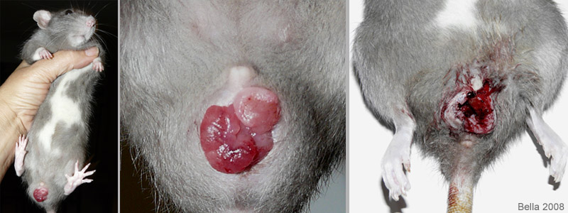 The first two photos were taken while Jango was still alive. The bulbous tissue coming from her vaginal opening is the cancer. In the last photo much of the tissue has been removed by laser, however the cancer went too deeply into the vagina to be operable. |
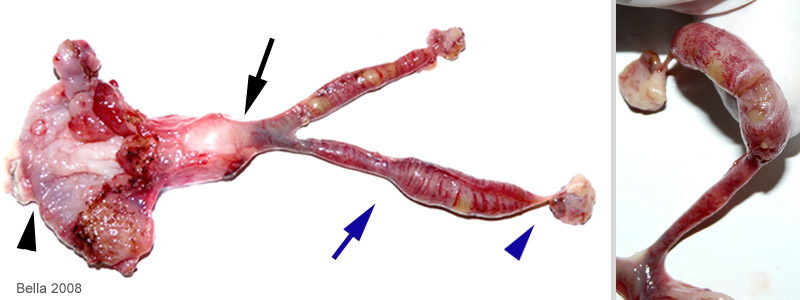 In the first photo we have removed the diseased reproductive tract. The black arrow head indicates the vagina/vaginal canal which is thickened and cancerous. The black arrow points to the uterine neck/cervix which also is thicker than normal. The blue arrow shows the left uterine horn. Both uterine horns have abnormalities in size, shape, and color. The blue arrow head points to the left ovary. In the second photo the left uterine horn has been cut open to reveal the cystic endometrial hyperplasia. The cells of the uterine wall (endometrium) have multiplied, the endometrial glands have become cystic with mucus, and mucus fills the lumen of the uterus. The white blood cells are indicative of uterine infection (pyometra). |
Necropsy and photos: J. “Bella” Hodges
Veterinary surgeon: R. McKinnis, DVM
Histopathology by IDEXX RADIL

