Figure 6: Exophytic squamous cell carcinoma: preputial mass in male rat (Remus).
Case history and photos
History
Remus was a pink-eyed-white (PEW) rescue, neutered male, 18-24 months of age with no previous health issues.
Clinical Signs
Three weeks before death the owner noticed 2 holes in the skin on the underside of the penis, as well as a swollen area and a putrid odor. Over the next few weeks (with treatment) the holes became larger, although there was no cutaneous necrosis or indication of healing process regarding the skin at the edge of the wound.
Over the course of disease Remus showed no decreased appetite or difficulty urinating.
Diagnosis
After ruling out both bacterial and fungal infections, it was determined to be some type of neoplasia of the preputial gland.
Treatment
Owner brought rat in for treatment a week following initial clinical signs.
Baytril and Clavamox were initiated, and an injection of dexamethasone was given (which did help to reduce the size of the mass to the left of the penis). The area was irrigated twice daily with sterile saline to remove debris and promote healing. Animax was topically applied to the wound, and buprenorphine was given for pain management.
With no evident signs of improvement, Baytril was continued while the Clavamox was stopped and an antifungal cream was used in conjunction with a hydrogel wound dressing ointment.
Outcome
Antibiotics helped to clear and control any secondary infection; however due to invasiveness and progression of the cancer it was decided to humanely euthanize.
Follow-up
A necropsy was performed, slides were gram stained, and tissue was sent to the IDEXX RADIL for histopathology.
MICROSCOPY
Gram stains showed abundant gram-negative rods, moderate filamentous gram-negative rods, and moderate gram-positive cocci.
NECROPSY
The mass was located attached to the length of the underside of the penis and did not appear to invade the penis. No preputial glands were noted. Several small subcutaneous nodules were located on the lower left quadrant of the abdomen. The liver had multiple dark areas similar in appearance to infarcts.
HISTOPATHOLOGY
SUMMARY
This animal had a preputial mass characterized as an exophytic squamous cell carcinoma.
Penis- no significant lesions
Preputial mass- Exophytic squamous cell carcinoma; focal severe exophytic proliferation of atypical squamous epithelium; these hyperplastic atypical epithelium forms tubular structures, intradermal finger like projections, sworls and multifocal nests of atypical epithelial cells scattered through the dermis; most of the tubular structures have moderate size lumens which are filled with moderate amounts of degenerate neutrophils, necrotic cellular debris, few dyskeratotic cells and moderate amounts of keratin; there is moderate to severe cellular atypia of the squamous epithelium characterized by severe anysokariosis and anysocytosis, the nuclear morphology of epithelial cells varies from round to oval, frequently with a vesicular appearance, with 1 to 3 prominent nucleoli; few areas show cells with scant cytoplasm and deeply basophilic nuclei resembling cells of the basal layer of the skin; mitotic figures are moderate and range from 0-2 per 400x power field; in adjacent skin, there is multifocal cellular swelling and dyskeratosis of the squamous epithelium with mild multifocal spongiosis; multifocally small to mid size keratin pearls are scattered though the squamous epithelium; there is mild multifocal parakeratotic hyperkeratosis and multiple epithelial erosions and ulcers; eroded and ulcerated areas are covered with an eosinophilic scab and contain moderate to severe suppurative infiltrate with superficial secondary bacterial colonization; some areas of the tumor are surrounded by a fibrous sheath of connective tissue that is formed by proliferation of fibroblasts and a mixed histiocytic plasmacytic infiltrate.
Necropsy Photos
Note: Arrowheads indicate the head of the penis. Arrows indicate the mass.
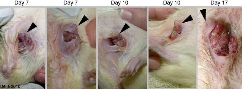 Remus was brought in for treatment on the seventh day after the owner first notice there was a problem. As you can see, in the photos marked “Day 7,” the entire area around the penis was extremely swollen. During irrigation a large amount of caseous debris was removed from the two openings on the the sheath. In the Day 10 photos the swelling around the area had decreased significantly. The skin receded to the point where all that was left was the fur on the head of the penis that was connected to the abdominal fur by a very thin strip of skin. The borders of this wound, and the lack of healing or necrosis, figured heavily in the diagnosis of a neoplasm. The odor was similar to the foul odor of badly infected/blocked preputial glands, hence our theory that this cancer was involved with those glands. In the Day 17 photo the mass is greatly enlarged, the skin has receded more, and new areas of tumor growth are noticeable. |
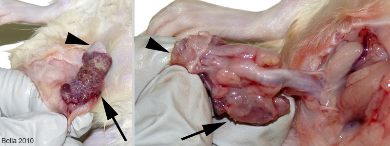 The above photos were taken during the necropsy. As the skin was removed the extensiveness of the mass could be seen. In the photo on the right the strip of fur, connecting the head of the penis to the abdominal fur, has been detached giving us a clear view of the actual penis. From this we can see that the preputial mass was wrapped about 3/4 of the way around the penis. |
Necropsy and photos by Joanne “Bella” Hodges
Microbiology by Kellie Blackwell
Histopathology by IDEXX RADIL
Case pathologist: Dr. Franklin Lopez
Consulting vet: Dr. Sharon Sernik, Westmonte Animal Clinic, Altamonte Springs, FL

