Fig. 3a: Necropsy of 10-month-old rat with megacolon (untreated)
Case history and photos
History
Argento, a 10-month-old male silver dumbo rat weighing 1.5 pounds. Shortly after fathering a litter, which produced 3 early onset megacolon afflicted babies, Argento began to bloat and was diagnosed with late onset aganglionic megacolon. It came on suddenly and intervention could not dislodge the blockage. Within a week of his initial bloating Argento became lethargic, the bloating increased substantially, his appetite diminished, and he refused to drink. At this point he was humanely euthanised.
Discussion
High risk markings
From looking at Argento it was not apparent that he was high risk for megacolon. He had no markings typical of “high whites” at risk. The only apparent marking was a small white head spot that was barely perceptible against his light silver fur.
After his offspring were born there were markings on them that led us to believe that Argento was most likely a blazed rat. Several of the offspring had very small blazes that only went up the nose about an eight of an inch. It is possible that due to his light color, particularly at the muzzle, that he may have had a slight unnoticeable blaze.
The Necropsy of Late Onset
The necropsy of a late onset rat who has not been treated differs greatly with the necropsy (refer to Fig. 3b) of a rat who has been under treatment for megacolon.
In the treated rat, the stool has been kept soft using a special diet, blockages have been removed by way of manipulation or enema, and medication has been given to help the colon function. The late onset rat who has not received treatment (due to the condition being hidden) will often show severe impaction, backed up softer fecal matter in the colon and caecum, as well as excessive air in the rest of the intestines. Extremely late onset is often unrecognized until the disease has progressed too far for treatment. The reason this happens may be due to the fact that as the descending colon and rectum begin to block and enlarge it is hidden beneath other organs, fat, and muscle. As the caecum begins to back up some bloating may begin to show. The severe bloating comes when the blockage is complete and the breakdown of the static bacteria produces an abnormal amount of air (gas). At this point the bloating becomes obvious to the naked eye.
The Necropsy
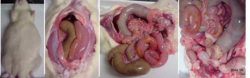
Description of above photos The photo on the above left shows Argento shortly after he was euthanized. You can see that the bloating is extreme. |
Description of above photos
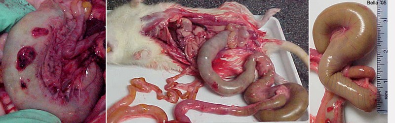
This row of photos begins with a close up of the ascending colon showing lesions where the bacterium has eaten through the wall of the colon. Once the bacteria breaks through the tissue and enters the abdominal cavity the rat then becomes septic. With most megacolon case this is presumed to be the final cause of death. |
Description of above photos
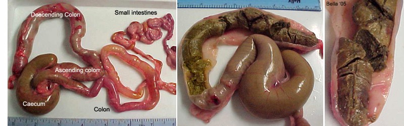
The first photo is another view of lower digestive system removed and tagged for reference. |
Necropsy and text by Joanne “Bella” Hodges
Photos courtesy of Bellaratta’s Nest Rattery

